Volume 4, Issue 11
November 2024
Innovations in Dental Imaging for Accurate Diagnosis and Treatment Planning in Canal Therapy
Manal Abdulrhaman Ajina, Mohammad Abdulrahman Algamdi, Nada Omer Balhaddad, Haifa Musa Alfaifi, Zamzam Abdullah Albahrani, Samirah Mousa Alamri
DOI: http://dx.doi.org/10.52533/JOHS.2024.41102
Keywords: Cone-Beam Computed Tomography, intraoral scanners, artificial intelligence, canal morphology, endodontic treatment
Advancements in dental imaging have dramatically transformed the field of endodontics, improving both diagnostic accuracy and treatment outcomes, particularly in canal therapy. Traditional two-dimensional radiography, while still widely used, is often limited in its ability to visualize complex root canal anatomies and periapical pathologies. Cone-Beam Computed Tomography (CBCT) has emerged as a superior alternative, offering detailed three-dimensional views of the teeth and surrounding structures. This technology allows clinicians to detect anatomical variations, root fractures, and resorptive defects with greater precision, which is critical for effective treatment planning. In addition to CBCT, the development of intraoral scanners (IOS) has enhanced the precision of dental impressions and treatment planning. IOS technology generates real-time digital images, eliminating the need for traditional impression materials and reducing errors. By providing highly accurate 3D models of the teeth and surrounding tissues, IOS improves the planning and execution of complex root canal procedures and restorations. Artificial intelligence (AI) has also begun to play a significant role in dental imaging, offering predictive diagnostic capabilities that complement traditional methods. AI-driven systems can detect subtle pathologies and analyze imaging data to predict treatment outcomes with increased reliability. This not only enhances diagnostic accuracy but also allows for more personalized treatment approaches. Together, these innovations in dental imaging are revolutionizing endodontic therapy by offering clinicians more precise diagnostic tools and improving patient care. As these technologies continue to evolve, their integration into routine clinical practice is expected to further enhance the accuracy, efficiency, and success of endodontic treatments. The adoption of advanced imaging technologies represents a major step forward in modern dentistry, particularly in the management of complex canal systems and challenging endodontic cases.
Introduction
Endodontic treatment, particularly root canal therapy, relies heavily on accurate diagnosis and precise treatment planning to ensure successful outcomes. A critical component of this process is dental imaging, which allows clinicians to visualize the complex internal structure of teeth and surrounding tissues. Over the years, advancements in dental imaging have revolutionized endodontic treatment, making it more efficient and predictable. Traditional two-dimensional (2D) radiographs, while still in use, have inherent limitations, particularly in detecting complex canal anatomies and assessing periapical pathologies. As a result, innovations in three-dimensional (3D) imaging technologies, such as Cone-Beam Computed Tomography (CBCT), have gained significant attention for their ability to provide more detailed and accurate diagnostic information.
The introduction of CBCT into dental practice has dramatically enhanced the ability of clinicians to identify root canal complexities, accessory canals, and variations in root anatomy, which are often missed in 2D radiographs. Studies have shown that CBCT offers superior diagnostic accuracy compared to conventional radiography, particularly in cases of re-treatment or previously failed endodontic procedures (1). In addition, CBCT enables the detection of periapical lesions and fractures that may not be visible on traditional X-rays, providing a more comprehensive assessment of the patient's condition (2). Another innovation in dental imaging is the use of intraoral scanners (IOS), which offer digital impressions for improved treatment planning and precision. IOS technology reduces the need for traditional impressions and allows for better visualization of the treatment area in real-time. This technology is particularly valuable in planning complex root canal therapies and evaluating the restoration of the tooth post-treatment (3).
Artificial intelligence (AI) is also emerging as a transformative tool in dental imaging, providing enhanced diagnostic capabilities through machine learning algorithms. AI-driven imaging systems can assist clinicians in identifying subtle signs of pathology and predicting treatment outcomes, further improving the accuracy and success of endodontic procedures (4). In this review, we will explore the innovations in dental imaging that have significantly contributed to accurate diagnosis and effective treatment planning in canal therapy.
Review
Recent advancements in dental imaging have greatly improved the accuracy of diagnosis and treatment planning in endodontic therapy. CBCT technology, for example, has significantly reduced the limitations associated with conventional 2D imaging. CBCT allows clinicians to visualize root canal systems in three dimensions, making it easier to identify root fractures, resorptive defects, and complex canal morphologies that are not easily detected by traditional radiography. This enhanced imaging capability provides a more detailed assessment, which is crucial for treatment planning and decision-making in complex cases (5). Moreover, the introduction of IOS has facilitated real-time imaging and digital impressions, minimizing errors associated with traditional impression techniques. By providing accurate 3D models of the teeth and surrounding structures, IOS has improved the precision of endodontic procedures and post-treatment restorations (6). The combination of these imaging technologies with AI further enhances diagnostic accuracy. AI-driven imaging systems, using machine learning algorithms, can detect subtle pathologies and predict treatment outcomes with increased reliability, making treatment planning more efficient and effective. These developments represent a shift towards more technologically integrated practices in modern endodontics, offering clinicians tools that not only improve diagnostic capabilities but also streamline complex procedures, ultimately enhancing patient care.
Advances in CBCT for Canal Morphology Assessment
CBCT has revolutionized endodontic diagnostics, particularly in the assessment of root canal morphology. Traditional two-dimensional radiography often falls short in visualizing the complex three-dimensional anatomy of the root canal system. CBCT, with its ability to produce high-resolution, three-dimensional images, provides clinicians with a comprehensive view of the internal structure of the teeth, making it invaluable for identifying complex canal configurations, accessory canals, and apical anatomy that are often missed in conventional radiographs (7).
One of the primary advantages of CBCT is its ability to assess canal morphology before treatment begins. Studies have shown that CBCT significantly improves the detection of root canal variations, such as C-shaped canals, which are particularly difficult to diagnose using traditional radiographs (8). This technology has also proven to be useful in cases where conventional imaging methods may produce distorted or overlapping images, such as in patients with multiple roots or those who have undergone prior endodontic treatment. In such cases, CBCT can reveal the intricate canal systems and help clinicians plan more precise and effective treatment strategies.
In addition to its diagnostic benefits, CBCT is also valuable in evaluating periapical lesions and other pathologies associated with root canal infections. For instance, it can detect periapical abscesses, granulomas, and cysts with greater accuracy than traditional radiographs. This is particularly crucial when determining the extent of infection and planning the necessary course of action, whether it be a root canal treatment or apical surgery. Furthermore, CBCT has proven beneficial in identifying vertical root fractures that might not be visible on 2D radiographs, providing a clear indication of whether the tooth is salvageable or requires extraction (9).
The role of CBCT extends beyond diagnosis, as it also enhances post-treatment evaluation. For example, clinicians can use CBCT to monitor the healing process after root canal therapy and detect any recurrence of infection. Moreover, CBCT allows for precise measurement of the length, shape, and curvature of root canals, which is crucial in determining the appropriate instrumentation and filling techniques during treatment. This high level of accuracy reduces the likelihood of treatment failure, thereby increasing the success rates of endodontic procedures. While the use of CBCT in endodontics has its benefits, it is important to consider the associated radiation exposure. Although CBCT emits lower radiation doses than conventional medical CT, it still delivers more radiation compared to standard dental radiographs. Therefore, clinicians must weigh the diagnostic benefits against the risks and ensure that CBCT is only used when necessary and justified (10).
The Role of Intraoral Scanners in Enhancing Precision during Canal Treatment
IOS have become an integral tool in modern dentistry, significantly improving the precision and efficiency of various dental procedures, including canal treatments. Traditionally, the process of diagnosing and treating root canals relied on manual impressions and radiographs, which can be time-consuming and prone to inaccuracies. IOS technology has changed this by allowing for the digital capture of high-resolution images of the patient’s teeth and oral structures, offering more accurate representations and aiding in the diagnosis and treatment of endodontic issues.
One of the primary benefits of IOS in canal treatment is its ability to create detailed 3D digital impressions of the tooth’s surface and structure. This eliminates the need for conventional impression materials, which can sometimes result in distortions. In the context of endodontic treatment, the accuracy provided by digital impressions allows clinicians to visualize the internal anatomy of the tooth more clearly and accurately assess the root canal morphology before commencing treatment (11). This is especially crucial in cases with complex canal anatomies, where precision is vital for successful outcomes. Moreover, IOS technology contributes to more efficient treatment planning. By generating real-time digital models, intraoral scanners allow clinicians to evaluate the condition of the tooth and surrounding tissues immediately. This helps in determining the most appropriate treatment approach, minimizing guesswork and reducing the potential for errors (12). The digital models produced by IOS also integrate seamlessly with computer-aided design/computer-aided manufacturing (CAD/CAM) systems, enabling the fabrication of highly accurate restorations, such as crowns or inlays, which are often required after root canal treatment.
Intraoral scanners also enhance patient comfort and reduce treatment time. Traditional impression methods can be uncomfortable for patients, particularly those with sensitive gag reflexes. IOS eliminates this discomfort by capturing digital impressions without the use of impression trays or materials. Furthermore, the immediacy of digital imaging reduces the number of clinical visits required, as the data can be instantly transmitted to dental laboratories for further processing or restoration fabrication (13, 14). Another advantage of IOS in endodontics is the improvement in communication between clinicians and patients. The high-quality digital images produced by intraoral scanners can be displayed in real time, allowing patients to better understand their dental condition and the proposed treatment plan. This visual aid enhances patient education and increases treatment acceptance, as patients can see the actual state of their teeth and the necessity for intervention.
Application of Artificial Intelligence in Dental Imaging for Predictive Diagnosis
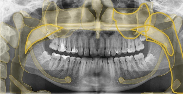
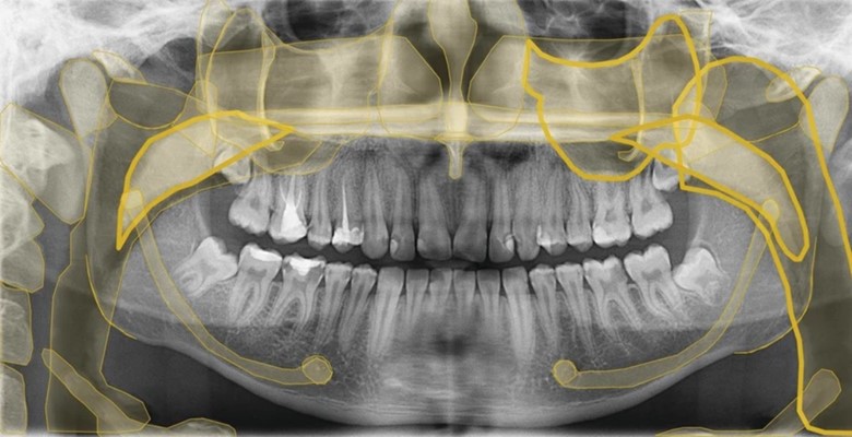
AI is rapidly transforming various medical fields, including dentistry, by enhancing diagnostic capabilities through machine learning and predictive algorithms. In dental imaging, AI applications are particularly valuable for improving the accuracy and efficiency of diagnosing complex conditions, such as those encountered in endodontic treatment. AI's ability to process large amounts of data and identify patterns in imaging has the potential to revolutionize how clinicians diagnose, plan treatments, and predict outcomes for canal therapy. One of the primary ways AI is being integrated into dental imaging is through the use of convolutional neural networks (CNNs), which are designed to analyze visual data with high precision. CNNs can automatically detect and segment dental structures, such as root canals, with a high degree of accuracy, reducing the risk of human error in identifying anatomical complexities or pathologies (15). This is particularly beneficial in diagnosing root canal infections, fractures, and other abnormalities that might not be immediately visible in conventional radiographs. By automating the detection process, AI can assist clinicians in making more accurate and timely diagnoses, ultimately improving treatment outcomes. For instance, some AI have been reported to outline the anatomical structure of the radiographic image, making it easier for dentists to keep track and easily differentiate between different structures on the radiology image (Figure 1) (16).

Figure 1: AI generated anatomical structures of a panoramic x-ray (16).
AI also enhances predictive diagnosis by utilizing data from past cases to forecast potential treatment outcomes. For example, AI algorithms can analyze a large dataset of previous endodontic treatments to predict the likelihood of treatment success based on specific imaging features. Factors such as canal morphology, periapical health, and pre-existing conditions are taken into account, allowing clinicians to make more informed decisions about treatment plans (17). This predictive capability is especially valuable in complex cases where the prognosis may be uncertain, as it helps clinicians assess potential risks and benefits before proceeding with treatment. AI-powered systems can be used to identify subtle signs of pathology that may be overlooked during manual assessments. For instance, AI can analyze CBCT images and detect minute changes in bone density or the presence of early-stage periapical lesions, which may not be immediately apparent to the human eye (18, 19). By identifying these early indicators, AI helps clinicians intervene sooner, potentially preventing the progression of disease and minimizing the need for more invasive treatments. AI's integration into dental imaging also contributes to more standardized diagnostic practices. Since AI systems rely on objective data rather than subjective interpretation, they can provide consistent diagnostic results across different practitioners and institutions. This standardization is particularly important in ensuring uniformity in treatment planning and improving overall patient care in endodontics.
Conclusion
Innovations in dental imaging, including CBCT, intraoral scanners, and AI, have significantly enhanced the accuracy of diagnosis and treatment planning in endodontic therapy. These technologies allow clinicians to visualize complex canal anatomies, predict treatment outcomes, and improve procedural precision. As these tools continue to evolve, their integration into routine dental practice will undoubtedly lead to better patient outcomes and more efficient care. The ongoing advancements in imaging will continue to shape the future of endodontics.
Disclosures
Author Contributions
The author has reviewed the final version to be published and agreed to be accountable for all aspects of the work.
Ethics Statement
Not applicable
Consent for publications
Not applicable
Data Availability
All data is provided within the manuscript.
Conflict of interest
The author declares no competing interest.
Funding
The author has declared that no financial support was received from any organization for the submitted work.
